Seaweeds of the South African South Coast
![]()
![]()
![]()
![]()
![]()
![]()
Order Ceramiales
Family Rhodomelaceae
Chondria sp. Indet.
Thalli erect, a few to 10 cm tall, main axes 500 - 1200 µm in diameter, sparsely branched, dark red to purplish, usually with striking patches or spots of green or turquoise iridescence. Holdfast of repent stoloniferous branchlets; erect axes single or several; axes and branches tapering basally and distally, branching polystichous, irregular, rather sparse. Apices with pits, usually containing trichoblasts. In cross-section, axial cells, five periaxial cells and another 1-2 layers of large cells clearly visible sections, surrounded by 2-3 (-4) layers of cells becoming smaller towards the outside. Cell dimensions rather variable, depending on individual specimens and part of thallus sectioned Axial cell 65-90 µm diameter; periaxial cells 100-200 µm diameter, usually ovate in cross section (elongated radially); next layer of cells the same size as periaxials or slightly larger; cell sizes then decreasing radially through 3-5 layers, outer (epidermal) layer of radially elongated cells 15-25 µm long and 10-15 µm wide. Tetrasporangia borne in short, clavate or spindle-shaped terminal branchlets that are often in clusters.
Collections, ecology and regional distribution
Distinctive on account of the iridescent spots, this species is recorded from De Hoop to Mkambathi (24-47). It is not uncommon in the lower eulittoral zone, where it often grows in taller turfs and in among other seaweeds such as Hypnea spicifera.
Note: This species looks very similar to C. scintillans Feldmann from southern Europe and the Mediterranean, but the whole genus requires molecular study.
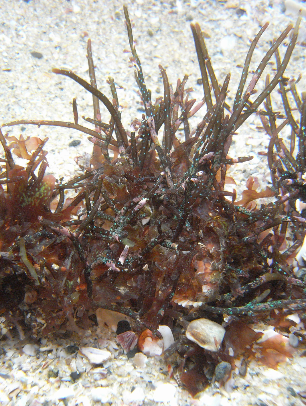
Chondria sp indet. Morgan Bay specimens, in situ, showing habit and iridescent turquoise spots (specimen D 2598).
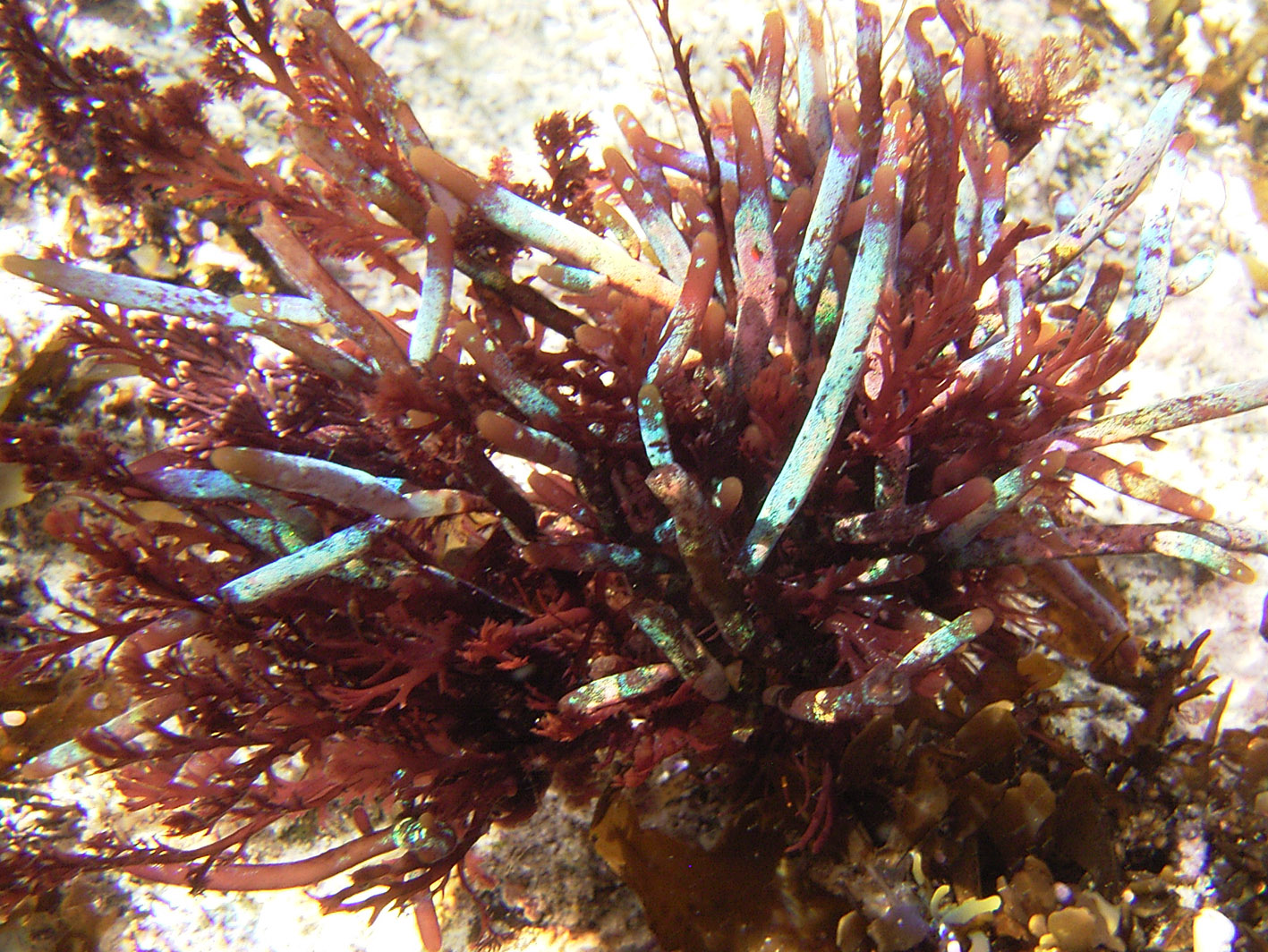
Chondria sp indet. Morgan Bay specimens - note iridescent turquoise spots (specimen D 2598).
Chondria sp indet. De Hoop specimens, in situ, showing patches of turquoise iridescence.
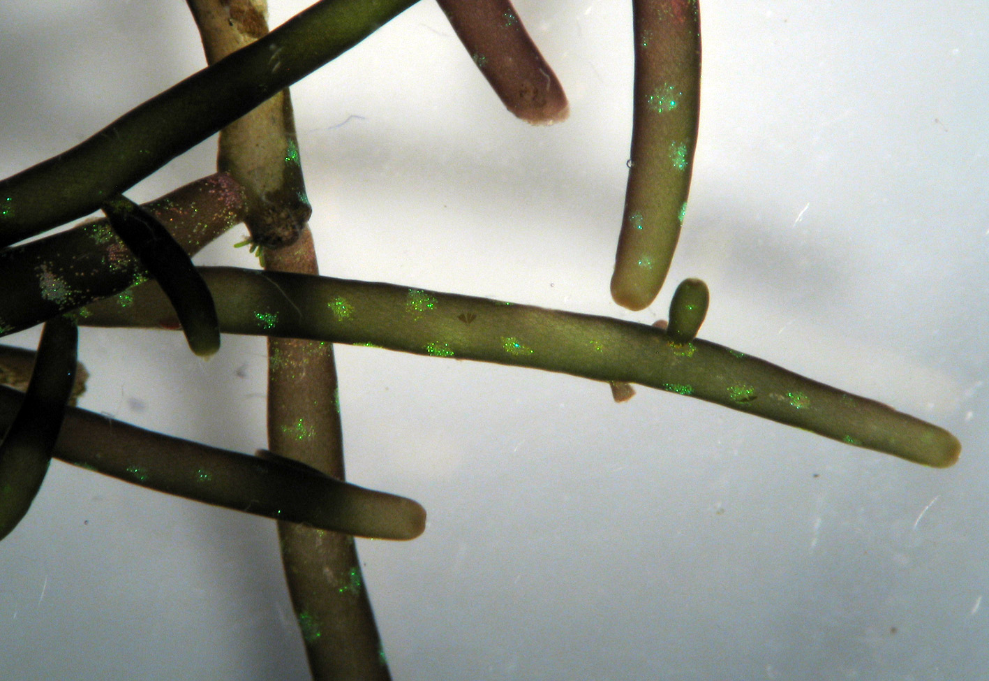
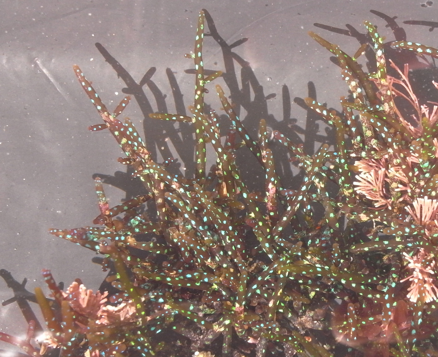
Chondria sp indet. Morgan Bay specimens, magnified to show iridescent turquoise spots and frond shape (specimen D 2598).
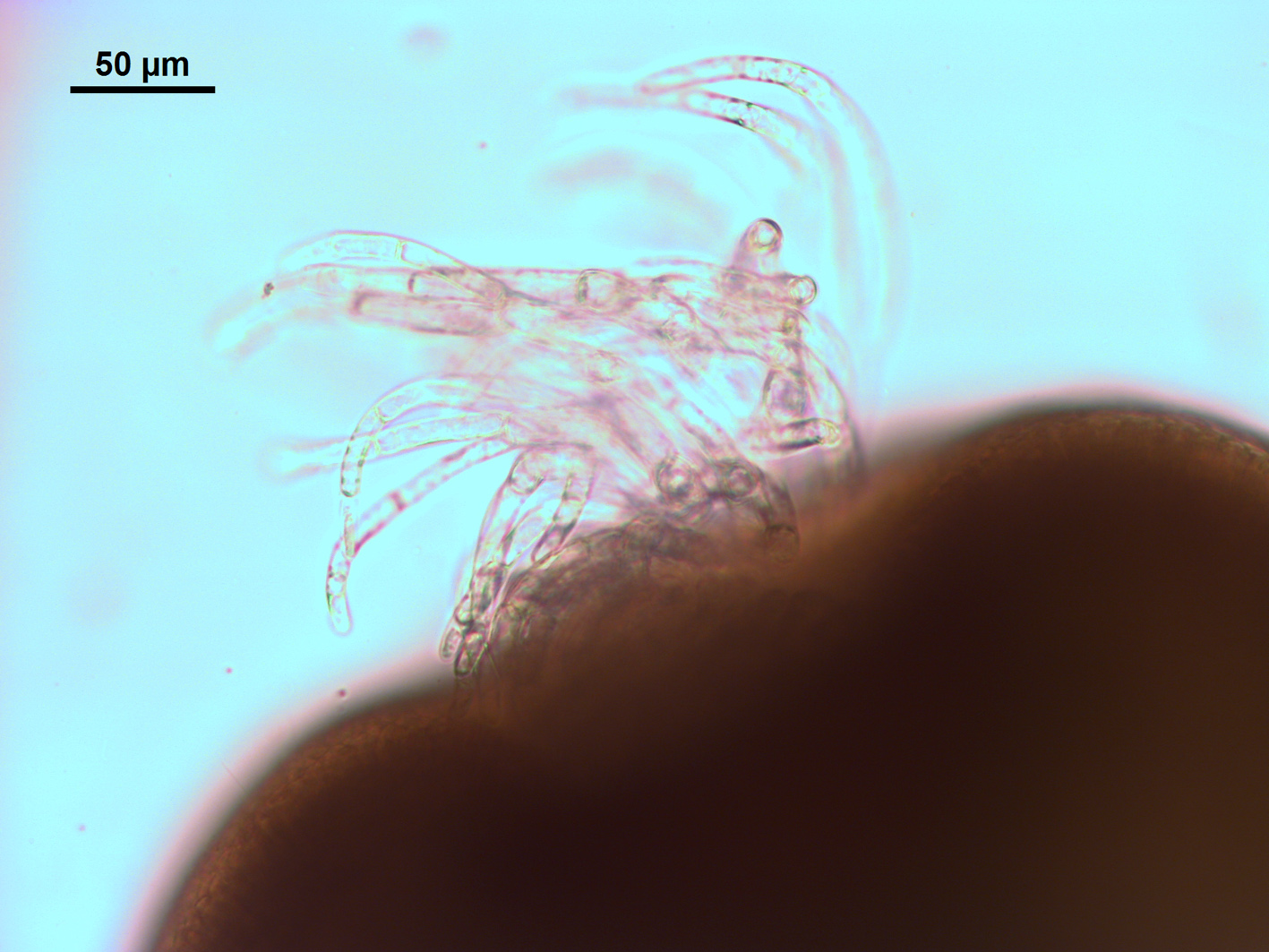
Chondria sp indet. Morgan Bay specimen showing apical pit with emergent trichoblasts (specimen D 2598).
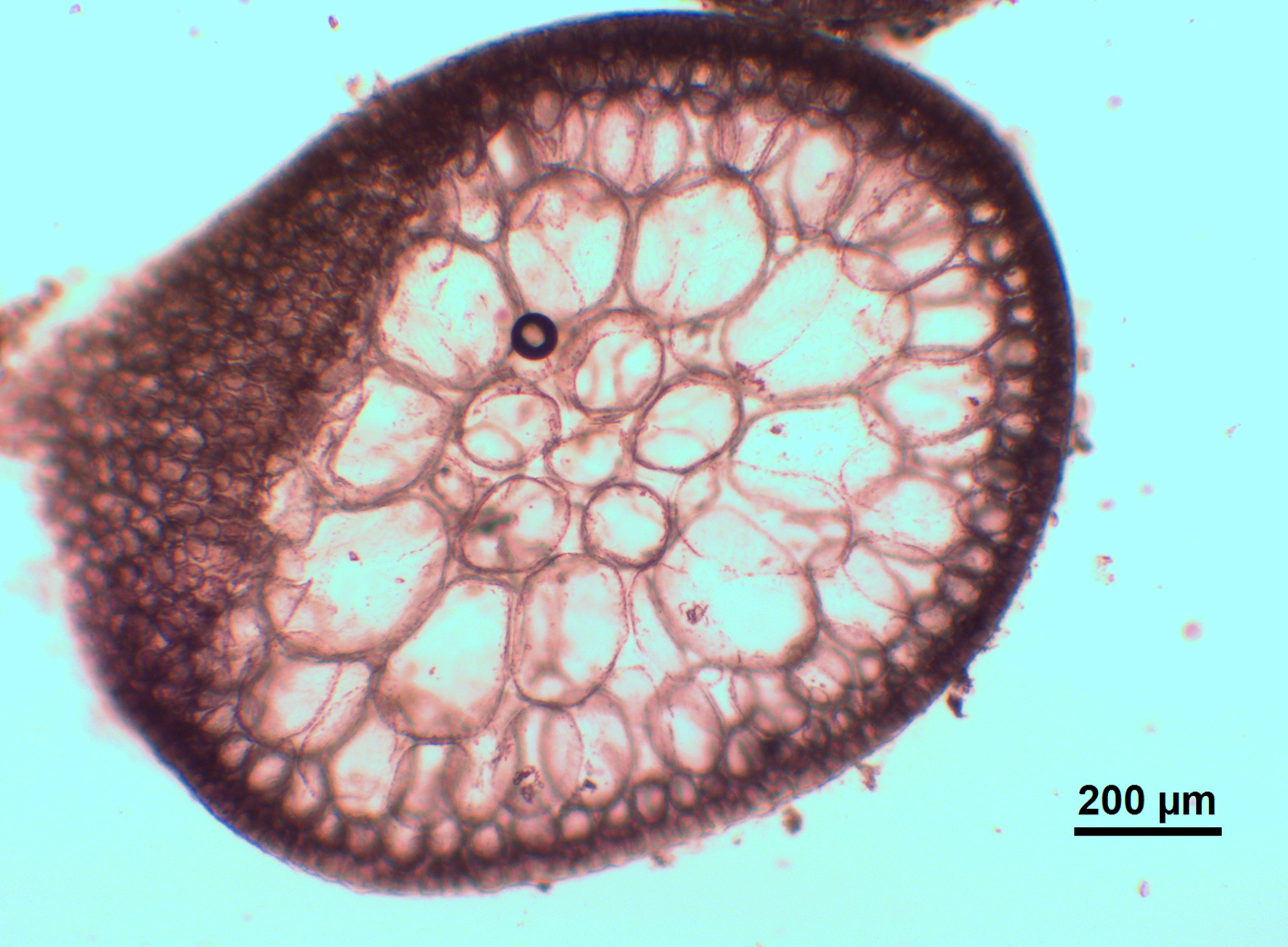
Chondria sp indet. Morgan Bay specimen – cross section showing 5 periaxial cells, etc. (specimen D 2598).
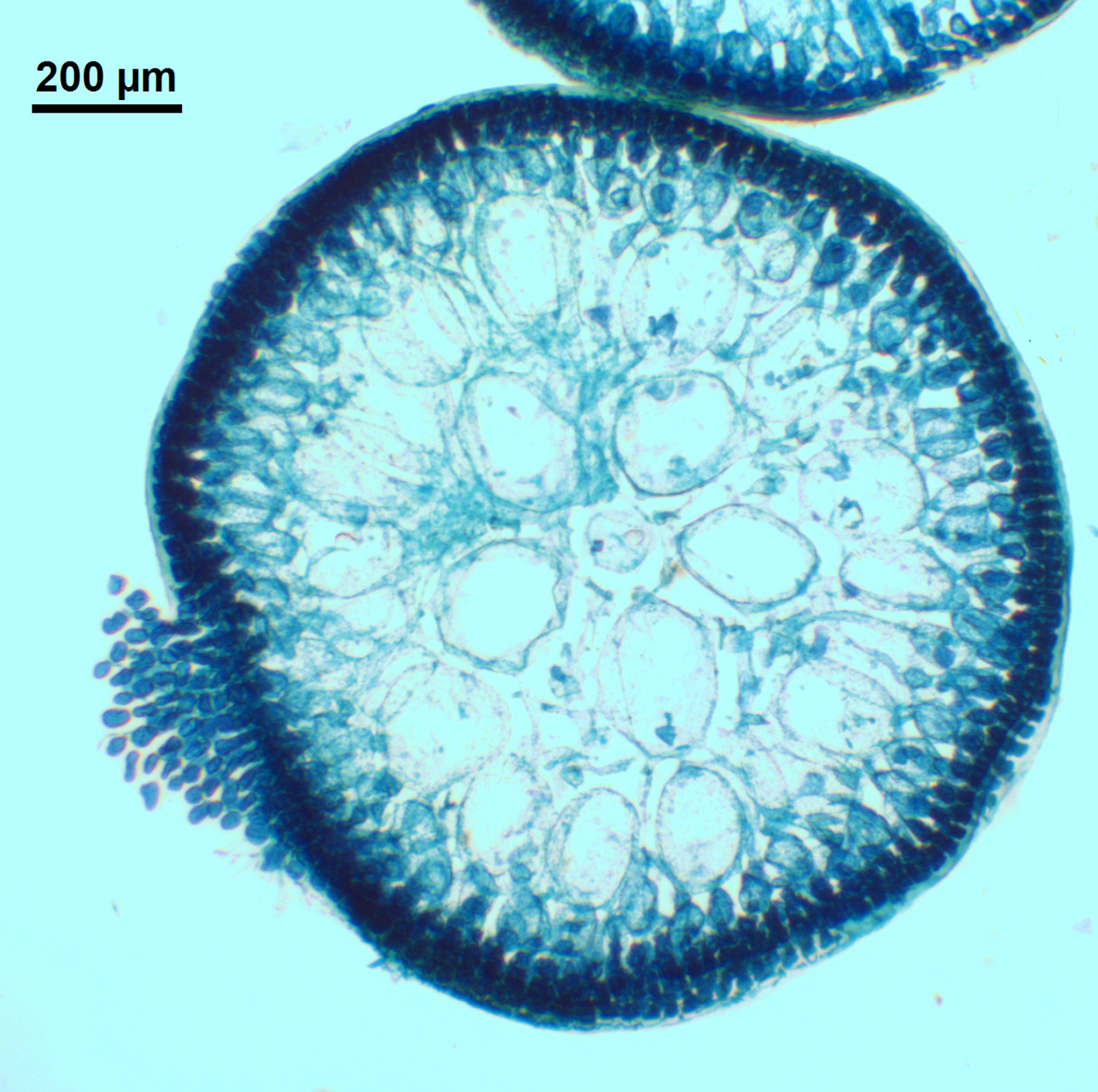
Chondria sp indet. East London specimen – cross section showing anatomy (stained slide EC 355).
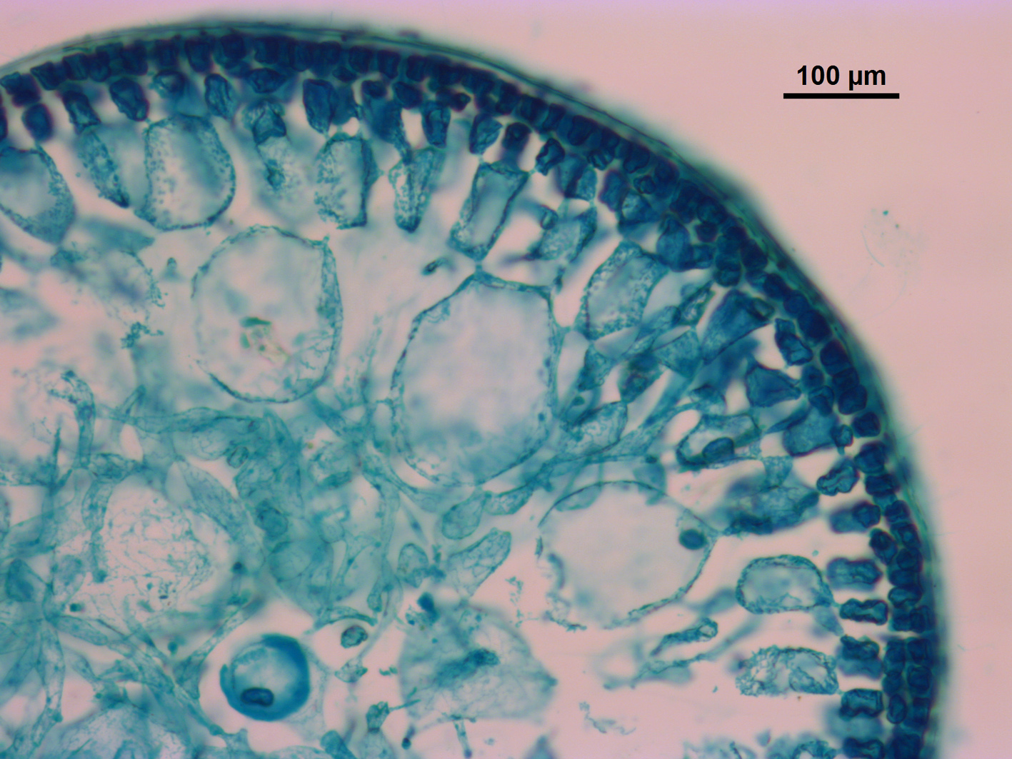
Chondria sp indet. East London specimen – cross section showing outer cortex and detail of anatomy (stained slide EC 355).

Chondria sp indet. Morgan Bay specimens, pressed mounts (specimen D 2598).
Cite this record as:
Anderson RJ, Stegenga H, Bolton JJ. 2016. Seaweeds of the South African South Coast.
World Wide Web electronic publication, University of Cape Town, http://southafrseaweeds.uct.ac.za; Accessed on 28 March 2026.