Seaweeds of the South African South Coast
![]()
![]()
![]()
![]()
![]()
![]()
Order Nemaliales
Family Scinaiaceae
Scinaia capensis (Setchell) Huisman 1985: 417
Plants pale to bright red, mucilaginous, up to 10 (-14) cm tall, somewhat bushy, with small discoid holdfast. Axes erect, cylindrical, dichotomously branched up to 10 times, about 2 mm diameter (3 mm wide in pressed specimens), with blunt apices. Structure multiaxial, with a narrow core of densely interwoven longitudinal filaments surrounded by a layer of fairly loose radiating filaments with very narrow cells lumens, and a solid epidermal layer. Epidermis comprising a mixture of chloroplast-containing cells (about 5-10 µm in diameter in surface view) and broader, thick-walled colourless vesicular cells (up to 25 µm diameter in surface view). Spermatangia superficial, cut off from narrow epidermal cells. Carposporophytes globose, immersed in the sub-epidermal layer. Tetrasporophytes unknown.
Collections, ecology and regional distribution
Recorded from Melkbosstrand on the west coast to Mzamba (15-47). Found in lower eulittoral pools, the eulittoral fringe, and in the sublittoral. Most common in spring and early summer, but sporadic in occurrence.
World distribution: South African endemic.
Type locality: Port Alfred (Silva et al. 1996).
Notes: Stegenga et al. (1997) point out that the tetrasporic phase, which is unknown, may be “acrochaetioid” in form (i.e. an open filament), similar to that known for S. furcellata (Turner) Bivona (Boillot 1968), and therefore inconspicuous. S. capensis was previously known as Pseudogloiophloea capensis (Setchell) Levring. S. capensis has also been collected from Meeuw Island in Saldanha Bay. There is one unusually large specimen of S. capensis (more than 30 cm long), collected from Kowie, in BOL.
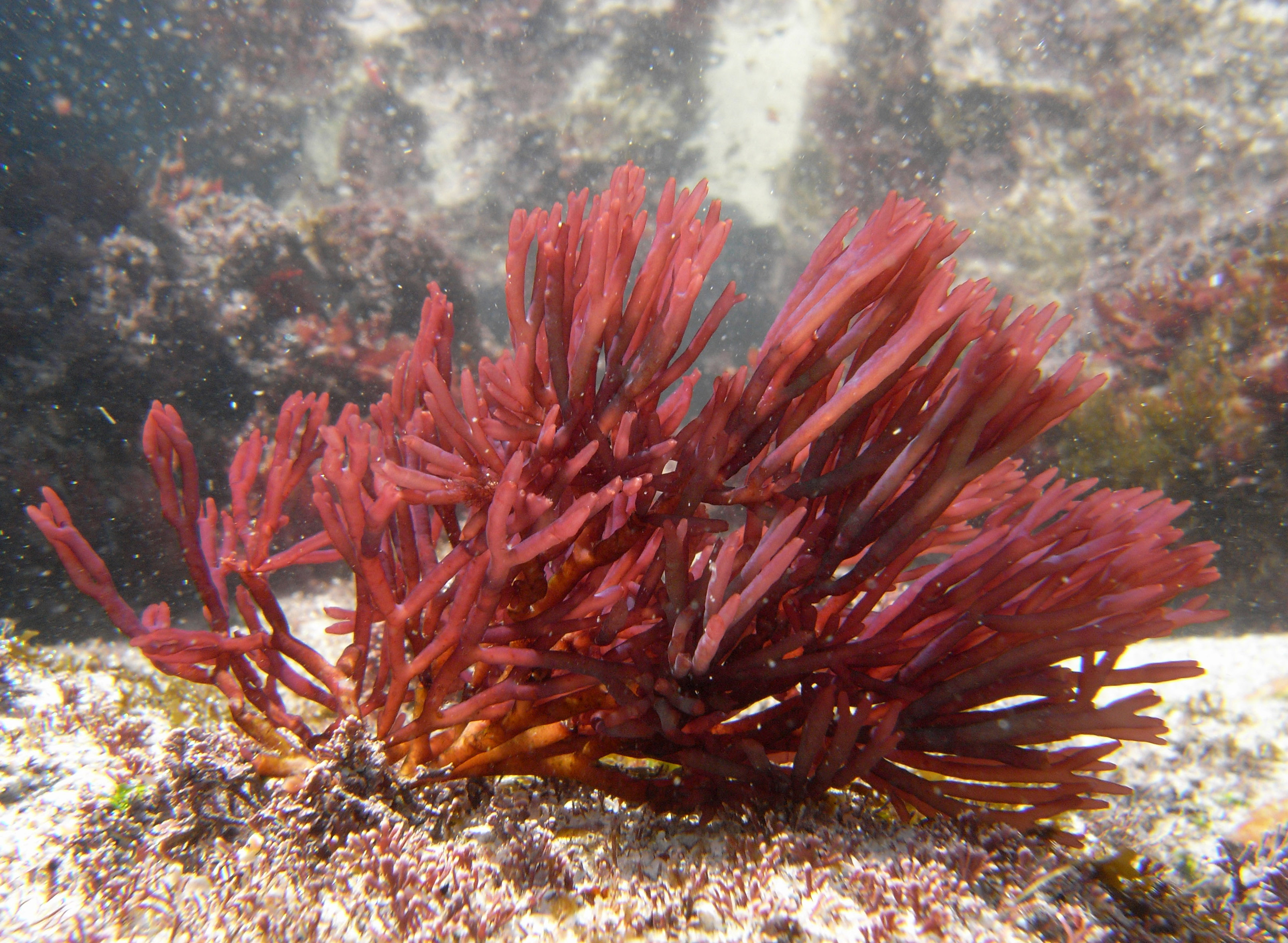
Scinaia capensis, underwater.
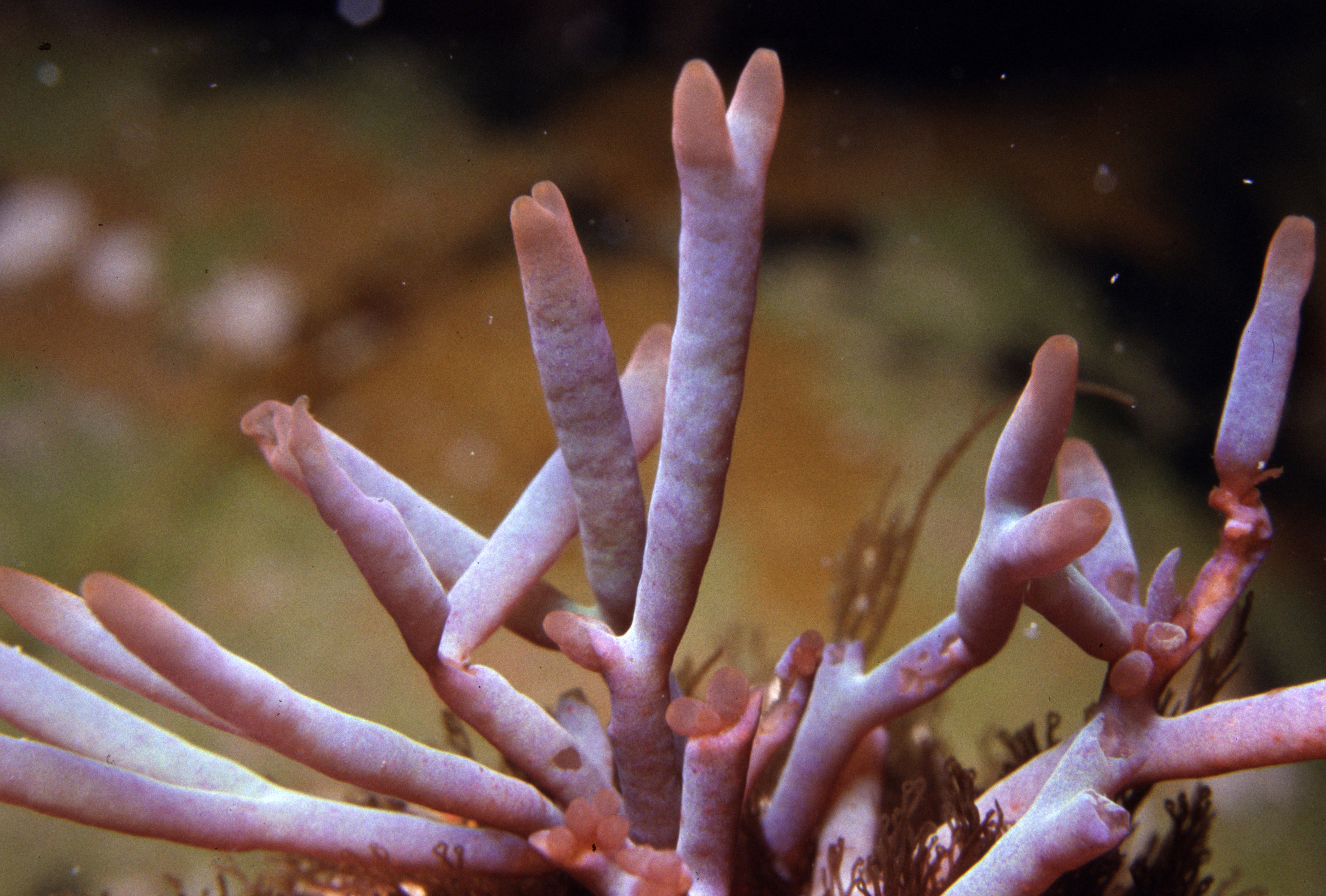
Scinaia capensis, underwater, showing bluish iridescence.
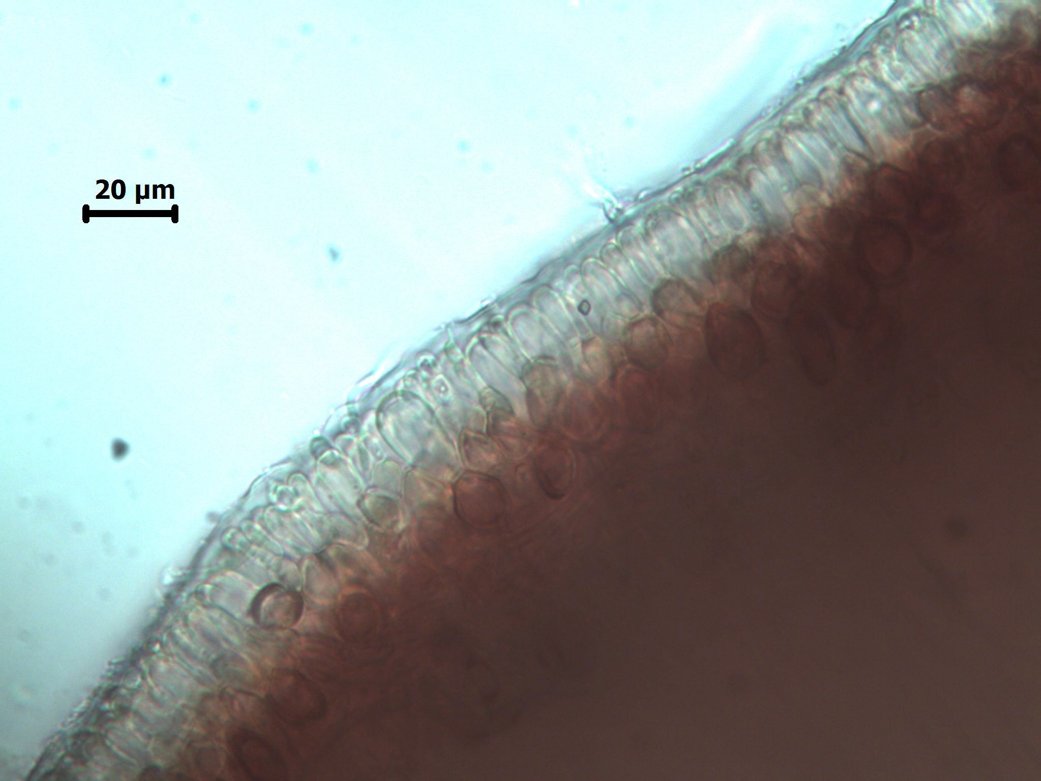
Scinaia capensis, cross-section showing faintly pigmented, narrow epidermal cells among larger colourless vesicular cells.
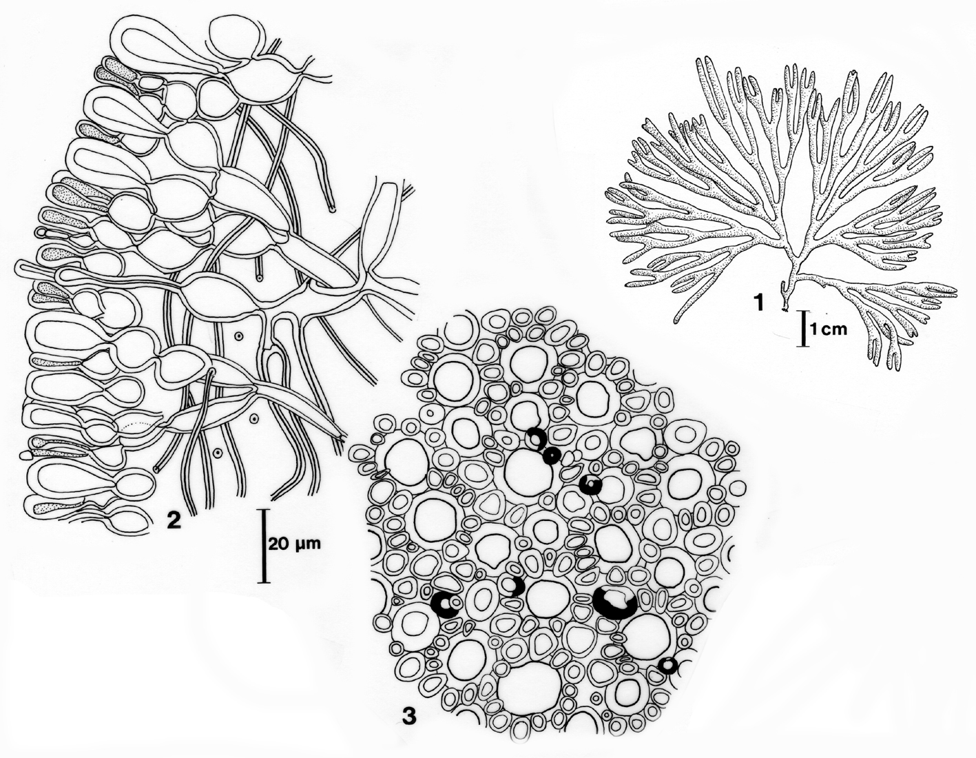
Scinaia capensis. 1. Habit. 2. Cross section of cortex. 3. Surface view of cortex. Reproduced from Stegenga et al (1997).
References Scinaia capensis
Boillot, 1968. Sur l’existence d’un tétrasporophyte dans le cycle de Scinaia furcellata (Turner) Bivona, Nemalionales. Compte Rendu de l’Academie des Sciences, Paris, 266: 1831-1832.
Huisman, J. M. 1985. The Scinaia assemblage (Galaxauraceae, Rhodophyta): a re-appraisal. Phycologia 24: 403-418.
Silva, P.C., Basson, P.W. & Moe, R.L. 1996. Catalogue of the benthic marine algae of the Indian Ocean. University of California Publications in Botany 79: 1-1259.
Stegenga, H., Bolton, J.J. and R. J. Anderson. 1997. Seaweeds of the South African west coast. Contributions from the Bolus Herbarium 18: 655 pp.
Cite this record as:
Anderson RJ, Stegenga H, Bolton JJ. 2016. Seaweeds of the South African South Coast.
World Wide Web electronic publication, University of Cape Town, http://southafrseaweeds.uct.ac.za; Accessed on 11 March 2026.